
Digital X-ray
- Skeleton
- Chest
- Intestinal tract
- KIDNEYS AND URINARY TRACT
- Veins
Digital X-ray
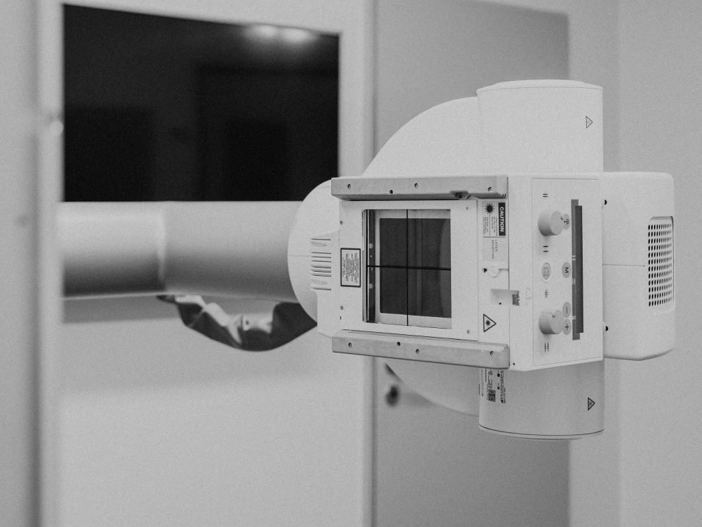
For many reasons, digital X-ray imaging is the preferred choice in modern medical diagnostics. It is non-invasive and painless, provides quick results, and the quality of its images is superior to those obtained with conventional X-ray imaging. Due to the high sensitivity of the digital detectors, the radiation dose is relatively low. In addition, digital X-ray images can be quickly forwarded to referring doctors and hospitals and archived without any loss of quality.
- SKELETAL X-RAYThis safe and pain-free type of examination provides valuable information concerning the condition of the bones and joints from head to toe. Major areas of application are imaging of the skull (paranasal sinus, mandibular joint); X-ray imaging of the entire spinal column, including specialised imaging for orthopaedic issues (full-leg imaging, functional imaging); as well as joint examinations of the upper extremities (shoulder girdle joints, shoulder joint, elbow joint and hand skeleton, including the wrist joints) and the lower extremities (pelvic X-ray, hip joint X-ray, knee joint X-ray and foot skeleton X-ray with ankle joints).
- CHEST X-RAY (THORACIC X-RAY)Thoracic radiography can quickly and easily provide valuable information concerning the condition of the heart, the lung and the chest (thorax) more generally. Amongst other things, it is used for the diagnosis of diseases and injuries and the evaluation of the condition of implants.
- DIGESTIVE TRACTDigital fluoroscopy provides detailed real-time images of all sections of the digestive tract, from the mouth to the large intestine. Depending on the requirements, any of the following methods may be used:
- Videofluoroscopic swallow study: The act of swallowing (deglutition) is observed in real time as the patient swallows liquid or solid contrast material.
- Abdominal X-ray: For the diagnosis of inflammations, ulcers and tumours, a contrast agent is administered. Patients are required to fast before the procedure, but are allowed to eat as soon as the examination has been completed.
- X-ray of the small intestine: In this procedure, the passage of a contrast agent through the small intestine is captured in a series of images that help in the diagnosis of strictures, tumours, inflammations, ulcers and other abnormalities.
- X-ray of the large intestine: This procedure, also known as irrigoscopy, is usually employed when other methods, such as colonoscopy, are not possible or additional information is required.
- KIDNEYS AND URINARY TRACT
Digital X-ray imaging of the kidneys and the urinary tract (urography) provides detailed information for the diagnosis, monitoring and assessment of a wide variety of diseases and conditions of the urinary tract, such as kidney stones. The examination itself is completely pain-free. - VEINSThe digital examination of veins (phlebography) is a valuable tool in the diagnosis and evaluation of venous disease, such as deep vein thrombosis (DVT), chronic venous insufficiency and venous malformations. Digital X-ray imaging may also be employed before an operation in order to visualise the anatomy of the veins. Digital examination of the veins is usually carried out for the lower extremities (legs), but in rare cases also for the upper extremities (arms).